AlRasheed Hospital Radiology Department:
Accurate Diagnosis with Advanced Technologies
The department is headed by a team of highly experienced doctors and radiologists, alongside highly trained radiology technicians and technologists. Our team is dedicated to providing exceptional quality services to patients, focusing on delivering accurate and rapid results to support effective treatment decisions
These devices and technologies include:
To ensure the highest standards of diagnosis, our Radiology Department is equipped with the latest international devices and technologies, including:
A digital X-ray, or digital radiography, is a modern form of X-ray imaging that uses digital sensors instead of traditional photographic film to capture images. The resulting images are available almost instantly on a computer, offering enhanced clarity and a lower radiation dose compared to conventional X-rays.
-In our department we have one Ceiling-Suspended System device and two mobile units device in addition to C-arm devices in operation theater and endoscopy units.
-The amount of radiation from a standard X-ray is very small and is often compared to the natural background radiation you receive from the environment over a few days.
-If you are or might be pregnant, you should inform your doctor. They may take extra precautions or suggest an alternative imaging test, such as an ultrasound.
A CT (computed tomography) scan is a medical imaging procedure that uses X-rays and a computer to create detailed, cross-sectional images of the inside of the body. It is used to help diagnose a wide range of conditions, detect abnormalities like tumors, and guide medical procedures. During the scan, you lie on a table that slides into a large, ring-shaped machine that rotates around you as it takes the images.
-It uses a series of X-ray images from different angles around your body. A computer then processes these images to create detailed cross-sectional “slices” of your bones, soft tissues, and blood vessels. It is also known as a CAT scan.
-The scan is fast and would take maximum of 5 minutes.
-CT angiography (CCTA), which uses a contrast dye to show blockages or narrowing in the arteries, and a coronary calcium scan, which identifies calcium deposits to help assess the risk of future heart attack
-The procedure is generally avoided during pregnancy unless absolutely necessary. Alternatives like ultrasound or MRI may be used to avoid radiation exposure to the fetus.
An ultrasound scan uses high-frequency sound waves to create images of the inside of the body, which can be used to diagnose medical conditions, monitor pregnancy, and guide procedures. A transducer (probe) is placed on the skin with a gel to transmit sound waves that bounce off internal structures, creating echoes that a computer turns into an image. This is generally a non-invasive and painless procedure, though it may cause some discomfort.
-Ultrasound scans do not use radiation and are considered safe because they use high-frequency sound waves to create images of the body, unlike X-rays and CT scans, which do use ionizing radiation. The sound waves in an ultrasound are inaudible and are reflected as echoes to form a real-time picture, and the procedure is generally painless, though some pressure may be felt.
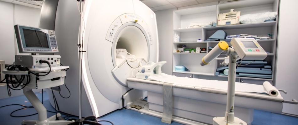
An MRI (magnetic resonance imaging) scan is a medical imaging technique that uses strong magnetic fields and radio waves to create detailed pictures of the inside of the body, such as organs, soft tissues, and bone. It is a non-invasive procedure that helps diagnose conditions, plan treatments, and assess their effectiveness. A person lies on a table inside a large, tube-shaped machine during the scan, which can take a while, and it is important to remain still.
-Deep learning and AI are transforming MRI scans by improving image quality, significantly reducing scan times, and enabling more accurate and faster diagnosis. They achieve this through techniques like deep learning reconstruction (DLR), which creates high-resolution images from less data, and by automating tasks such as patient positioning, which saves time and reduces variability. AI algorithms can also enhance diagnostic accuracy by detecting complex patterns in images that are difficult for the human eye to see.
For fast and reliable results, and for more information about our services, please visit AlRasheed Radiology department or contact us today.
+96264777444 | +962790777444